


Dental Radiographs
|
 Full Mouth Periapicals
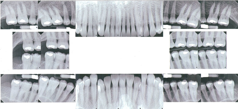 |
|
|
|
Panographs
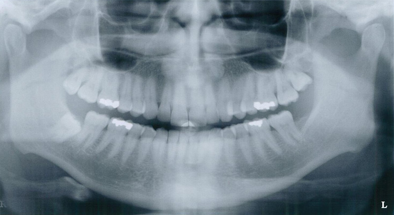
A common screening film is the panograph, which gives an overview of both upper and lower arches and the surrounding anatomic structures. When taking this x-ray no film is placed in the patient's mouth, and the x-ray machine circles around the head. While a useful screening tool, and important for implant placement, it lacks the detail needed for a complete periodontal exam.
Click the x-ray for a larger view.
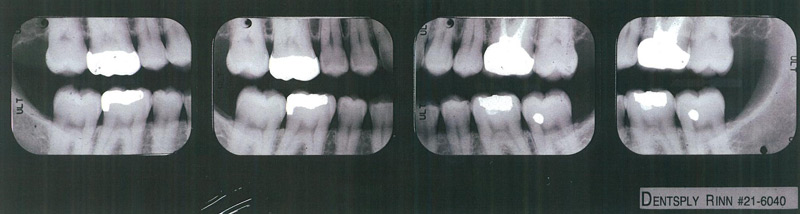
Bitewings
The bitewing x-ray is commonly used to detect decay, and may be used in the general dentist's screening in conjunction with the panograph. It shows the crowns of the upper and lower teeth at the same time, and shows good detail. Normally 4 films are taken. Unfortunately, the bone is often not visible, which limits the usefulness of bitewings in periodontics. Bitewings are generally recommended every 6-12 months to check for decay.
Click the x-rays for a larger view.
 |
|
|
|
|
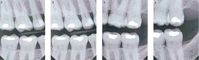
Vertical Bitewings
Vertical bitewings are films that show both upper and lower teeth, but the view extends farther down the root, generally giving an excellent image of the bone. Normally 4-7 views are taken. In cases of severe bone breakdown the film will not show all the bone, and root canal lesions also do not show. They are taken in place of traditional bitewings.
Click the x-rays for a larger view.
 |
|
|
|
Digital X-Rays
Digital x-rays don't use conventional x-ray film, but rather the image is stored in a computer and projected on a monitor. There are a number of advantages to digital x-rays, including even less radiation needed for exposure, and having the ability to manipulate the image on the computer to optimize the view. The major drawback is when other dentists need copies of the x-rays. While printing the digital x-ray on paper is reasonably accurate, many practitioners find the printed results don't have the diagnostic quality of normal x-rays, or of the digital x-ray as seen on the monitor. If the second dentist also uses digital x-rays there is no problem, as images can be sent on floppy disk or by e-mail. However, at this time only about 10% of dentists use digital, and because we send an original copy of all films to the general dentists, at present regular x-rays are still necessary.